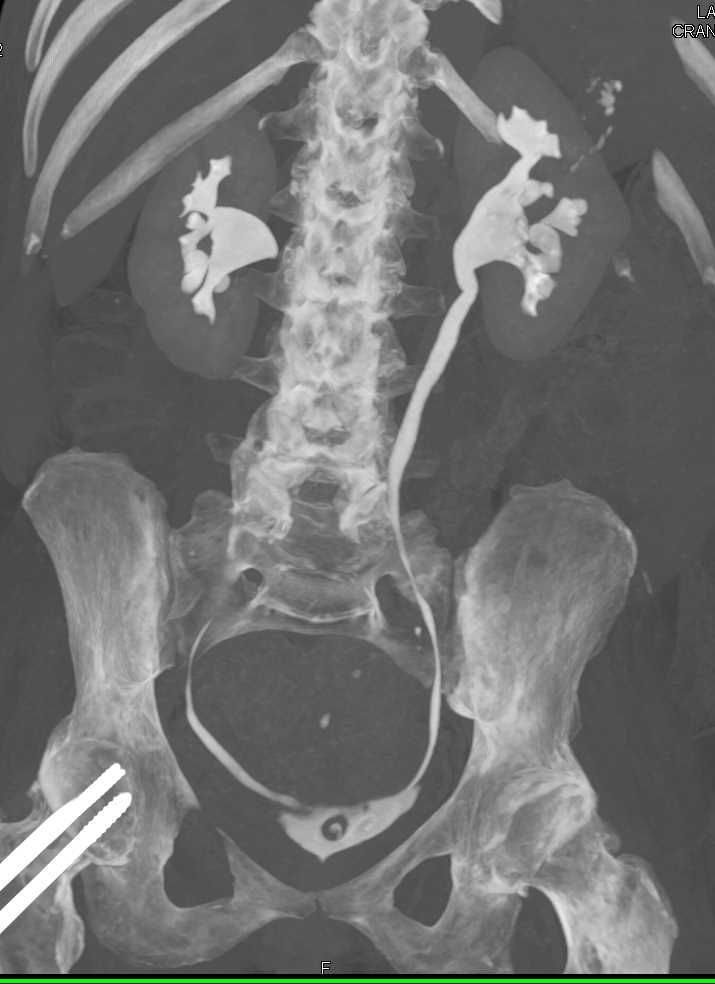

Staghorn stones composed of struvite are usually associated with urinary tract infection caused by a urea-splitting organism, the most common of which is Proteus mirabilis. Most staghorn stones are composed of mixtures of magnesium ammonium phosphate and calcium carbonate apatite, but cystine and uric acid stones can also present in this fashion.

4 These stones usually fill the renal pelvis and branch into several or all of the calices. Staghorn calculi are branched stones that occupy a major part of the renal collecting system. Staghorn stones are slightly more common in females. Males and those with a family history of renal stones are more likely to be affected. 1,2 Without preventative intervention, up to 50% of patients have a recurrence within 5 years after successful treatment. Urolithiasis affects about 10% to 12% of the population worldwide, and the incidence is increasing. Stone analysis revealed that the calculi were composed of 76% magnesium ammonium phosphate (struvite) and 24% calcium carbonate( apatite. Left PCNL was also performed with clearance of all the residual stones ( Figure 1D). Secondstage right PCNL for the residual stones was performed 2 weeks later, and all the stones were cleared ( Figure 1C). Simultaneous right percutaneous nephrolithotomy (PCNL) was performed, and a ureteral stent was inserted at the end of the procedure ( Figure 1B). The patient underwent transurethral incision of the right ureterocele and ureteroscopic removal of the ureteral stone. Serum calcium, phosphate, urate, blood urea( nitrogen, creatinine, and electrolyte levels were normal. A radioisotopic study showed the differential function of the right and left kidneys to be 44% and 56%, respectively, with scarring in the upper pole of the right kidney. A subsequent intravenous urogram (IVU) confirmed the presence of bilateral renal staghorn calculi and a right ureteral stone within a ureterocele. Plain radiography with a kidney-ureter-bladder (KUB) view revealed multiple opacities in the region of both kidneys and specifically in a location that corresponded to each renal pelvis ( Figure 1A). Urinalysis showed 5 to 10 white blood cells and 3 to 5 red blood cells per high-power field. Other than mild loin tenderness, physical findings were normal. Her history included 3 urinary tract infections a Proteus species was cultured on each occasion. She did not have urinary frequency, urgency, or dysuria. Complete stone clearance is achieved after the third PCNL ( D).Ī 48-year-old woman sought medical attention after an episode of gross hematuria associated with mild right-sided loin discomfort. Treatment results are shown after the first percutaneous nephrolithotomy (PCNL) and removal of the ureteral stone ( B) and after the second PCNL ( C). Figure 1 – A pretreatment plain radiograph reveals bilateral renal stones and a right lower ureteral stone inside an ureterocele ( A).


 0 kommentar(er)
